
Biography Ueli Raz
| This is an introductory manual for a software you may be interested in, called Segmenter Dicom Viewer. After you have made CTs and MRIs, you get a CD by the hospital with an included program that hides more than it would show you what it should. The german Segmenter Dicom Viewer shows you the regular pictures in the three axis, together with all the pictures in 3D - through the body as it was scaned in every possible position you want. - Dicom ist the worldwide name of digital files out of x-ray, ct and pet-machines. | Ici, il y a un petit manuel d'introduction pour un logiciel qui pourrait vous intéresser, appelé Segmenter Dicom Viewer. Après avoir fait des CT et MRI, vous obtenez un CD de l'hôpital avec un logiciel inclus qui cache plus qu'il ne vous montrerait ce qu'il devrait. Le Segmenter Dicom Viewer allemand vous montre les images régulières dans les trois axes, avec toutes les images en 3D - à travers le corps comme il a été scanné dans toutes les positions possibles. - Dicom est le nom mondial des fichiers numériques sur les machines à rayons X, CT et MRI. |
=======> Software: https://www.dornheim-segmenter.com/produkte/dicom-viewer/ <=======
1 month free, afterwards ca. 20 Euro / $
Une mois gratuit, après 20 Euro (si vous voulez le programme)
Deutsch & english
You cannot
save pictures in this software, but you can make screenprints.
All the
pictures here are screenprints, edited in photoshop.
Vous ne pouvez
pas enregistrer des images dans ce logiciel, mais vous pouvez faire des
screenprints.
Toutes les images ici sont des screenprints, éditées dans
photoshop.
Alle Bilder auf dieser Seite: Screenprints.

1
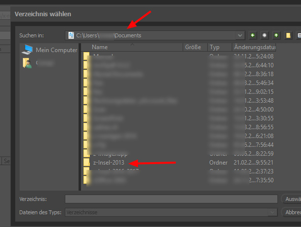
Après copier tout le CD du hôpital sur l'ordinateur, vous ouvrez avec "Ändern".
2

3

4

5

6
rouge = bon // purple = pas bon
Links auf den grünen Haken achten, rechts auf die Anzahl Bilder in einer Serie.

7
8
9
10
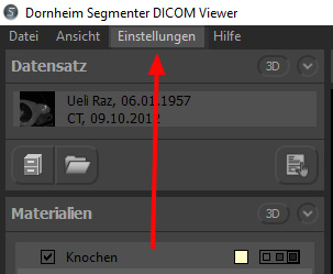
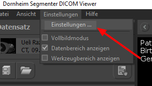
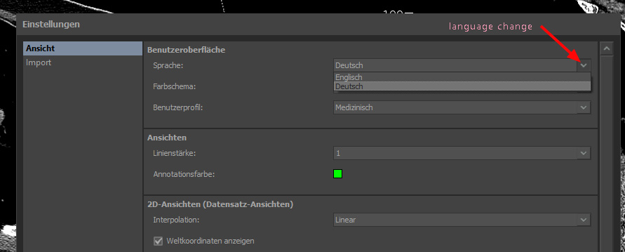
Seulement les langues allemand et anglais sont disponibles.

11

12
Souris gauche:
rotation // Souris droite: contraste (comme ça, vous ouvrez le corps...)
Linke Maustaste: drehen, mit ctrl: verschieben, rechte Maustaste:
Kontraständerunng (weiche Partien verschwinden)

13
Die Schnittebenen lassen sich verschieben.
-------------------------End of manual----------------------
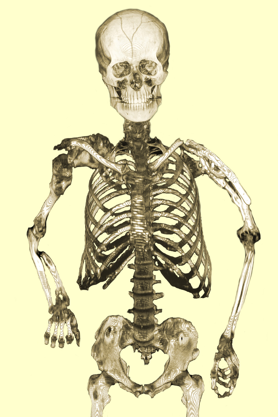
Collage Photoshop:
1) Schädel 2012
mit Segmenter Dicom Viewer
2 ) Kopf und Hals 2012 mit Segmenter Dicom Viewer
3) Abdomen, Schulter, Arme und Becken 2014 mit Segmenter Dicom Viewer
4)
Beide Ellenbogen 2014 mit RadiAnt Dicom Viewer
Fiji
PET-CT-Viewer gibt es auch noch:
Fiji is free, but difficult.
Fiji est gratuit,
mais difficil.
Fiji ist open source, also gratis, aber schwierig.
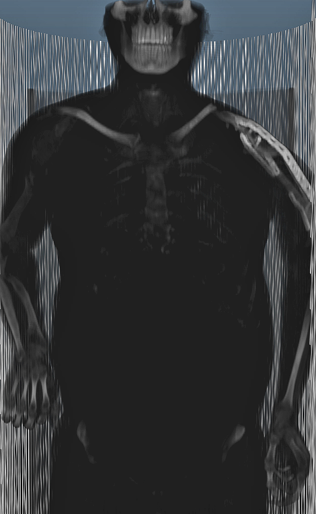
2014
(the
classical Ollier is on the right side, the cancerdevil on the left ... so far)
(Ollier normal chez moi est le côté droit, mais le diable du cancer est à
gauche ... jusqu'à maintenant)
(Ollier sollte einseitig sein, bei mir rechts,
die mutierenden Chondrome sind aber links, bis jetzt...)
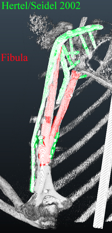
2012 = 2002
(toujours mon épaule gauche)
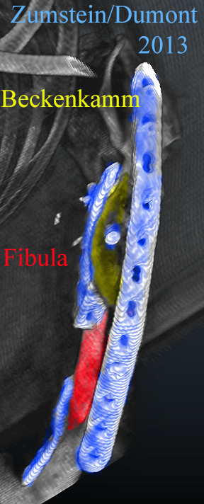

2013
 |
 |
| 2012 | Chondrosarkom von aussen,
2012 Zeit des Wachstums: 10 Monate (Das Baumgeäst als Widerschein des Tumors, die ungeöffneten Weinflaschen zur Ablenkung.) |

2012 with chondrosarcoma 2014 without chondrosarcoma
 |
 |
| Juli 2002 - Januar 2013 | 2013 - |
 |
 |
| Juli 2002 - Januar 2013 |
2013 - |
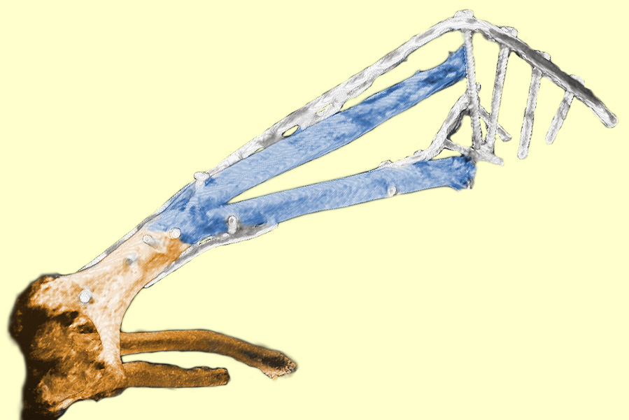
| Linke Schulter von hinten,
Ellenbogen angewinkelt Blau Fibula |
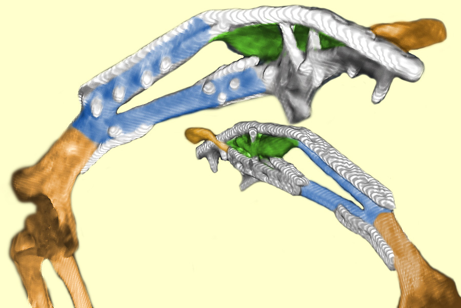
Linke Schulter von hinten, Ellenbogen
gestreckt, kleines rechtes Bild von vorne Blau Fibula gekürzt, grün Beckenkamm |

ur II
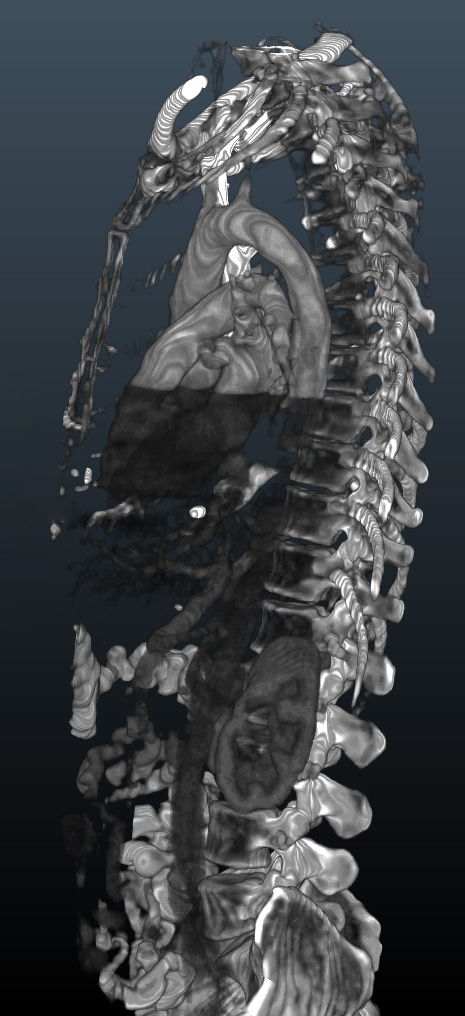
My heart

First I was
shocked about the emptiness of my head, then astonished about the pearl in the
brain...
D'abord, j'étais choqué du vide dans ma tête, puis étonné de la perle
au centre du cerveau...
(verkalkte Zirbeldrüse / "Verkalkung der Glandula pinealis")